Vibrational exciton nanoimaging of phases and domains in porphyrin nanocrystals
| Reviews and Highlights | Quantum Science | Molecular and Soft-matter | Ultrafast Nano-optics and Nanophotonics | Mineralogy and Geochemistry |
|---|
Eric A. Muller, Thomas P. Gray, Zhou Zhou, Xinbin Cheng, Omar Khatib, Hans A. Bechtel, and Markus B. Raschke
PNAS 6, 7030 (2020).
DOI PDF SI

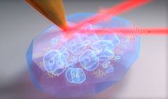
Much of the electronic transport, photophysical, or biological functions of molecular materials emerge from intermolecular interactions and associated nanoscale structure and morphology. However, competing phases, defects, and disorder give rise to confinement and many-body localization of the associated wavefunction, disturbing the performance of the material. Here, we employ vibrational excitons as a sensitive local probe of intermolecular coupling in hyperspectral infrared scattering scanning near-field optical microscopy (IR s-SNOM) with complementary small-angle X-ray scattering to map multiscale structure from molecular coupling to long-range order. In the model organic electronic material octaethyl porphyrin ruthenium(II) carbonyl (RuOEP), we observe the evolution of competing ordered and disordered phases, in nucleation, growth, and ripening of porphyrin nanocrystals. From measurement of vibrational exciton delocalization, we identify coexistence of ordered and disordered phases in RuOEP that extend down to the molecular scale. Even when reaching a high degree of macroscopic crystallinity, we identify significant local disorder with correlation lengths of only a few nanometers. This minimally invasive approach of vibrational exciton nanospectroscopy and -imaging is generally applicable to provide the molecular-level insight into photoresponse and energy transport in organic photovoltaics, electronics, or proteins.
News & Highlights: JILA
