
Imaging nanoscale heterogeneity in ultrathin biomimetic and biological crystals
| Reviews and Highlights | Quantum Science | Molecular and Soft-matter | Ultrafast Nano-optics and Nanophotonics | Mineralogy and Geochemistry |
|---|
Brian T. O’Callahan, Kevin T. Crampton, Irina V. Novikova, Tengyue Jian, Chun-Long Chen, James E. Evans, Markus B. Raschke, Patrick Z. El-Khoury, and A. Scott Lea
J. Phys. Chem. C 122, 24891 (2018).
DOI PDF SI
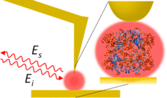
The mechanisms of interface biochemistry are often obscured through ensemble averaging of complex networks of proteins that exhibit varying degrees of structural heterogeneity. Here, we perform nanometer spatially resolved chemical spectroscopy using infrared vibrational scattering-scanning near-field optical microscopy (IR s-SNOM) to image protein–protein interactions and conformational heterogeneity of (i) a two-dimensional lipid-like peptoid membrane and (ii) a three-dimensional catalase crystal. In the peptoid membrane, which consists of stacked ∼4 nm high peptoid layers, spatio-spectral line width analyses reveal heterogeneity of the vibrational dynamics due to peptoid–substrate and peptoid–peptoid interactions. In s-SNOM imaging of catalase crystals, our results revealed complex secondary structures that vary over nanometer length scales. Broadening of vibrational resonances, as observed from the dehydrated catalase crystals, indicates a higher degree of heterogeneity as compared to the synthetic peptoid membranes. Our results thus demonstrate the utility of IR nanoscopy for the investigation of structural heterogeneity within biomimetic and biological systems with high spatio-spectral resolution and sensitivity.