
Hyper-spectral Raman imaging correlating chemical substitution and crystallinity in biogenic hydroxyapatite: Dentin and enamel in normal and hypoplastic human teeth
| Reviews and Highlights | Quantum Science | Molecular and Soft-matter | Ultrafast Nano-optics and Nanophotonics | Mineralogy and Geochemistry |
|---|
Ping Wang, Evan J.D. Anderson, Eric A. Muller, Fuhua Gao, Yisi Zhong, and Markus B. Raschke
J. Raman Spectrosc. 49, 1559 (2018).
DOI PDF SI
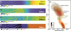
Micro‐Raman imaging and spectroscopy has become an established technique for the characterization of biogenic hydroxyapatite as, for example, the primary constituent of human teeth. However, few studies have yet gone beyond a qualitative analysis of the Raman response providing only limited insight into spatial heterogeneity of composition, structure, and degree of crystallinity. Here, we show how correlative electron microprobe and extended hyperspectral Raman imaging with high spatial and spectral resolution, with peak position and linewidth analysis, and from the μm to mm scale, provides insight into structural characteristics in dentin and enamel. From comparison of healthy and hypoplastic teeth as a representative tooth disease example, we determine variations in degree of crystallinity, both locally across the dentin?enamel junction, and with distinct long‐range spatial variations. We identify a correlation of spectral peak position and linewidth as a measure of crystal lattice disorder across tubules, dentin, dentin?enamel junction, and enamel. This correlative Raman imaging and analysis approach may help to provide a better understanding of apatite geochemistry and biomineralization.